Their location is variable but there is usually one just below and one about 10cm above the medial malleolus, and another one a little below the middle of the leg. Along the medial side of the calf behind the medial border of the tibia a variable number of perforating (anastomotic) veins connect the great saphenous with deep veins of the calf ( Fig. 113) for here it has most of its deep connections. This is an important part of the course of the great saphenous vein (see p. The great saphenous vein and the saphenous nerve lie in the fat, accompanied by numerous lymphatic vessels which pass up from the foot to the vertical group of superficial inguinal nodes. The subcutaneous surface of the tibia has subcutaneous fat in direct contact with the periosteum the deep fascia here is blended with the periosteum.
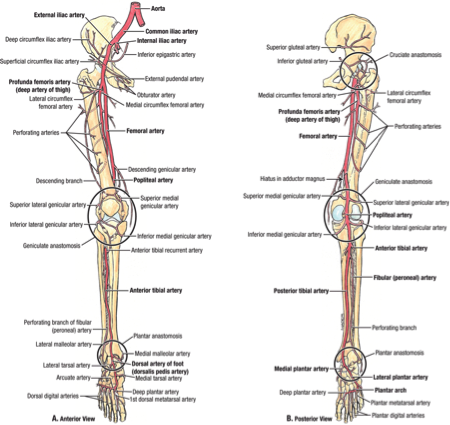
COMPARTMENTS OF LEG VASCULATURE SKIN
The lateral cutaneous nerve of the calf, a branch of the common peroneal, supplies deep fascia and skin over the upper parts of the extensor and peroneal compartments and the superficial peroneal nerve replaces it over the rest of these surfaces. The main nerve (anterior branch) often extends on the medial side of the foot as far as the bunion region: the first metatarsophalangeal joint. It usually bifurcates above the malleolus, and the branches run in front of and behind the vein.

The saphenous nerve gives off its infrapatellar branch, to supply the subcutaneous periosteum of the upper end of the tibia and the overlying and adjacent prepatellar skin it then descends just behind the great saphenous vein with which it passes in front of the medial malleolus. The cutaneous nerves are derived from the femoral nerve over the tibia and from the common peroneal nerve over the extensor compartment ( Fig. The front of the leg includes the subcutaneous surface of the tibia on the medial side and the extensor muscular compartment on the anterolateral side.

Last's Anatomy: Regional and Applied Part six.


 0 kommentar(er)
